

Exosomes are small membrane vesicles (30-150nm) that contain complex RNA and proteins. Nowadays, exosomes specifically refer to disc-shaped vesicles with a diameter of 40-100nm. All cultured cell types can secrete exosomes, which are naturally present in body fluids, including blood, saliva, urine, cerebrospinal fluid, and milk.
01.What are exosomes
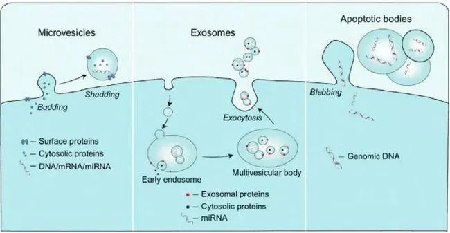
Exosomes are a type of extracellular vesicles. International Society for extracellular vesicles (ISEV) defines extracellular vesicles (EVs) as: "Cells secrete into extracellular lipid membrane-coated vesicles. There are three types of Apoptosis bodies, including Microvesicles, Exosomes and apoptosis bodies, according to their generation process, release pathway, size, content and function."
Microvesicle is an extracellular vesicle formed by outward budding or "extruding" of cell, containing cell membrane and some cytosolic components. The particle size of MVs ranges from 100nm to 1000nm, and the surface contains CD40, integrin, selectin and other proteins, and the content of phosphatidyl serine is high.
Apoptotic bodies are vesicles formed by cells that are "shed" or "broken" and released into the outer cell during apoptosis or death, which can be understood as "membrane structures that are gradually shed from the cell". Its morphological heterogeneity is the largest, the size of 50nm-5000nm, but generally speaking, the large particle size accounts for a higher proportion. The membrane usually contains more phosphatidyl serine.
The exosome originated from the inward budding of the cell membrane to form endosome, which can be discharged from the cell by exocytosis after fusion with the cell membrane through some processes such as early endosome, polycyst complex, directional assembly, and migration. Therefore, the composition and structure are more complex, and even contain some components "ingested" from the extracellular stroma or culture medium. Exosomes are about 50-150nm in size and contain conventional four-transmembrane proteins of cell membranes (CD9, CD63, CD81, etc.). Heat shock protein (Hsc70), lysosomal protein (Lamp2b), tumor sensitive gene 101 (Tsg101) fusion protein (CD9, flotillin, annexin) and other proteins, the membrane cholesterol and diacylglycerol content is high.
All three types of extracellular vesicles are derived from cells (the cells that give rise to extracellular vesicles are often referred to as their "mother cells" for convenience of description). Exosomes, microvesicles and apoptotic bodies can be generated in the same cell at different times and growth states. The three kinds of extracellular vesicles inevitably contain the components of the mother cell, such as membrane proteins, skeletal proteins, cytoplasmic proteins, phospholipids, metabolites, etc. Both its molecular composition and particle size are more or less overlapping.
Although the three types of extracellular vesicles are obviously different in theory, there is currently no technology to separate exosomes from microvesicles or apoptotic bodies, and no clear molecular markers to identify the three types of extracellular vesicles. They are often referred to as "extracellular vesicles".
Among the three kinds of extracellular vesicles, exosomes have the smallest average particle size, the highest average degree [the narrowest distribution range of particle size], the most complex composition and the most diverse functions. Therefore, the theory and application value is the highest, has been the most in-depth research and extensive application. Therefore, the mention of extracellular vesicles mostly refers to exosomes, which are often mixed with extracellular vesicles (EVs) in academic papers and daily communication.
02.Where do exosomes come from
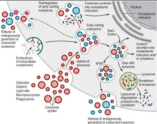
The origin, synthesis and secretion of exosomes go through the following processes: The cell membrane of the mother cell can form early endosome through endocytosis or "inward budding", and gradually mature into late endosomes and multivesicular bodies in the cell. MVBs). The rudiments of exosomes exist in MVBs in the form of intraluminal vesicles (ILVs). Subsequently, MVBs fuse with the cell membrane and expel ILVs out of the cell by exocytosis to become exosomes.
The maturation of early and late endosomes into polycystic bodies is also the "assembly process" of exosomes: A variety of intracytoplasmic substances including proteins (enzyme molecules, heat shock proteins, etc.), nucleic acids (mainly RNA such as mRNA, miRNA, piRNA, snoRNA, snRNA, rRNA, tRNA, Y-RNA, scRNA, etc.), metabolites and other active or passive loading into the intracellular body membrane or cyst.
Microvesicle is an extracellular vesicle formed by outward budding or "extruding" of cell, containing cell membrane and some cytosolic components. The particle size of MVs ranges from 100nm to 1000nm, and the surface contains CD40, integrin, selectin and other proteins, and the content of phosphatidyl serine is high.
Apoptotic bodies are vesicles formed by cells that are "shed" or "broken" and released into the outer cell during apoptosis or death, which can be understood as "membrane structures that are gradually shed from the cell". Its morphological heterogeneity is the largest, the size of 50nm-5000nm, but generally speaking, the large particle size accounts for a higher proportion. The membrane usually contains more phosphatidyl serine.
The exosome originated from the inward budding of the cell membrane to form endosome, which can be discharged from the cell by exocytosis after fusion with the cell membrane through some processes such as early endosome, polycyst complex, directional assembly, and migration. Therefore, the composition and structure are more complex, and even contain some components "ingested" from the extracellular stroma or culture medium. Exosomes are about 50-150nm in size and contain conventional four-transmembrane proteins of cell membranes (CD9, CD63, CD81, etc.). Heat shock protein (Hsc70), lysosomal protein (Lamp2b), tumor sensitive gene 101 (Tsg101) fusion protein (CD9, flotillin, annexin) and other proteins, the membrane cholesterol and diacylglycerol content is high.
All three types of extracellular vesicles are derived from cells (the cells that give rise to extracellular vesicles are often referred to as their "mother cells" for convenience of description). Exosomes, microvesicles and apoptotic bodies can be generated in the same cell at different times and growth states. The three kinds of extracellular vesicles inevitably contain the components of the mother cell, such as membrane proteins, skeletal proteins, cytoplasmic proteins, phospholipids, metabolites, etc. Both its molecular composition and particle size are more or less overlapping.
Although the three types of extracellular vesicles are obviously different in theory, there is currently no technology to separate exosomes from microvesicles or apoptotic bodies, and no clear molecular markers to identify the three types of extracellular vesicles. They are often referred to as "extracellular vesicles".
Among the three kinds of extracellular vesicles, exosomes have the smallest average particle size, the highest average degree [the narrowest distribution range of particle size], the most complex composition and the most diverse functions. Therefore, the theory and application value is the highest, has been the most in-depth research and extensive application. Therefore, the mention of extracellular vesicles mostly refers to exosomes, which are often mixed with extracellular vesicles (EVs) in academic papers and daily communication.
03.Where do exosomes go
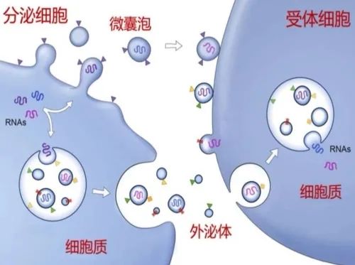
Compared with the relatively clear "linear process" of exosome generation, the whereabouts of exosomes after being released by cells is obviously complicated, changeable and full of uncertainty. If adjacent tissue cells can recognize and "capture" exosomes, exosomes are taken up and utilized by adjacent cells. If nearby cells cannot recognize and capture exosomes, exosomes will be transported to distant cells or tissues through the circulatory system.
The uptake and utilization of exosomes by nearby or distant tissue cells are mainly through the following different mechanisms of action:
① Exosome membrane surface ligands bind to receptors on the receiving cell membrane to activate receptor-mediated signal transduction pathways, and activated recipient cells ingestion of contents into cells through endocytosis.
② The receiving cells directly ingest exosomes into the cells by endocytosis, and the exosome contents are released into the cells. Some exosome components participate in the new multivesicular biosynthesis process.
③ Exosome membrane directly fuses with cell membrane, releasing exosome contents into the cytoplasm.
The whereabouts of exosomes is highly uncertain and influenced by many factors. There are directional migration with a clear destination, such as receptor-mediated specific binding and "homing" of stem cells or tumor cells. There are also aimless or random walks. It may be nonspecific phagocytosed by the circulatory system and reticuloendothelial system, or it may be changed by the local microenvironment and cell state of recipient cells.
04.What do exosomes look like
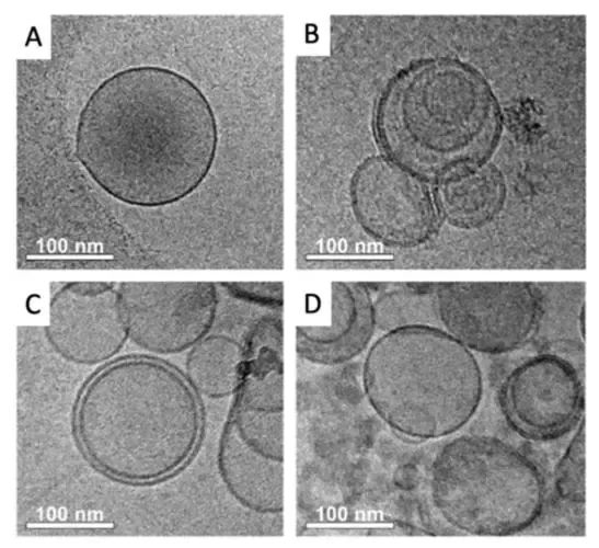
Because exosomes are too small to be observed by general microscopy, the morphology of exosomes can only be observed by electron microscopy. The first exosome images observed and reported by transmission electron microscopy (TEM) are such "cup-shaped" [FIG. 1]. Since then, it has been widely accepted and spread, and the morphology of exosomes is described as "cup-shaped". Since the sample processing before electron microscopy can cause the exosomes to "dehydrate" and "shrink", this generally accepted exosome morphology may be caused by the treatment rather than its natural shape.
Later, scanning electron microscopy (SEM) and cryo-electron microscopy (CEM) were used to observe and record exosomes with different shapes. More suggests that exosomes are "spherical in different sizes (Figure 2), with a double shell (Figure 3)". Not only the morphology of exosomes is revealed more clearly, but also the structure of exosomes is suggested.
外泌体的结构与组成
05.Structure and composition of exosomes
The structure of exosome is a vesicle structure. The exosome membrane [outer shell] is composed of a bilayer of phospholipids and proteins, and the interior is the so-called "cyst cavity". The exosome membrane has a hydrophobic structure, while the lumen is a hydrophilic environment. (Think of it as a "pissing cow ball," in which oil (phospholipids) and meat (protein) are blended into the shell, and the liquid inside is the exosomal "contents.")
To make Q-balls and meatballs, you need to know the types and combinations of oil and meat. Exosomes are as hard and elastic as bovine meatballs, and the hardness and elasticity of exosomes are mainly determined by the type and proportion of phospholipids on the membrane.
Exosome membranes are mainly composed of phospholipids and proteins.


 川公网安备 51010702003112号
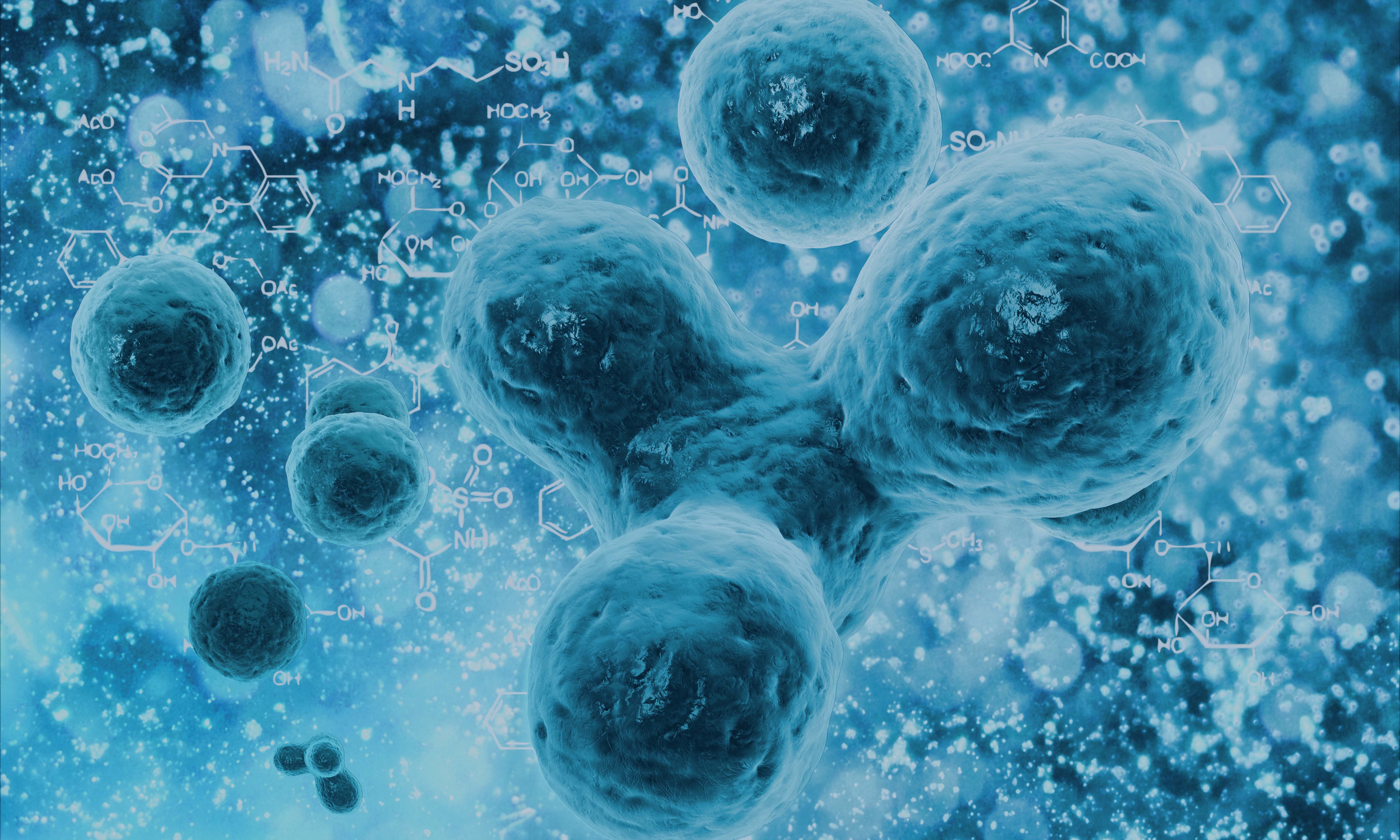 The difference between stem cells and immune cells